



Ultrasound-Guided Embryo Transfer: Does Better Visualization Improve IVF Outcomes?
Embryo transfer is one of the final and most technically important steps in in vitro fertilization (IVF). Even when high-quality embryos are available, the way they are introduced into the uterine cavity may influence the likelihood of successful implantation and pregnancy.
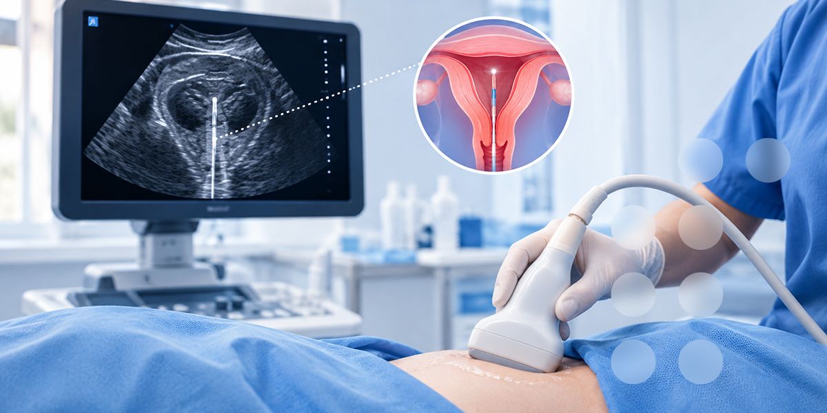
Ultrasound-guided embryo transfer allows clinicians to visualize the uterus and monitor the transfer catheter during the procedure. Compared with relying on tactile feedback alone, this approach provides real-time anatomical information that can help clinicians navigate the cervical canal, position the catheter, and deposit the embryo at an appropriate location.
But does improved visualization translate into better IVF outcomes? Evidence from randomized controlled trials, systematic reviews, and clinical practice guidance suggests that transabdominal ultrasound guidance can improve clinical pregnancy rates and may increase live-birth rates compared with the traditional clinical-touch technique. The size of the benefit varies across studies, however, and ultrasound guidance cannot overcome every factor that affects implantation.
This article examines the evidence, explains the practical steps involved, and outlines how fertility clinics can incorporate ultrasound guidance into a standardized embryo transfer protocol.
What Is Ultrasound-Guided Embryo Transfer?
Ultrasound-guided embryo transfer is a technique in which ultrasound imaging is used to monitor the placement of an embryo transfer catheter inside the uterus during IVF or other assisted reproductive technology (ART) procedures.
In conventional clinical-touch embryo transfer, the clinician relies primarily on tactile feedback, knowledge of uterine anatomy, and previously assessed uterine measurements to guide the catheter. Although experienced clinicians can perform transfers successfully using this approach, the exact catheter position may be less directly observable.
With ultrasound guidance, the clinician can assess the uterine cavity and observe the catheter's progress, helping to identify the intended deposition site and avoid unnecessary contact with the uterine fundus.
Two main approaches are used:
-
Transabdominal ultrasound guidance
A transabdominal probe is placed on the patient's lower abdomen. The resulting images help the clinician visualize the uterus and catheter during transfer. A moderately full bladder is commonly required to improve the acoustic window and may also help straighten the uterocervical angle.
This is the best-studied approach and is recommended by the American Society for Reproductive Medicine (ASRM) to improve clinical pregnancy and live-birth rates.
-
Transvaginal ultrasound guidance
A transvaginal probe is used to visualize the uterus from within the vagina. This approach may be useful in selected clinical circumstances, including when transabdominal visualization is difficult or bladder filling is poorly tolerated.
However, the evidence comparing transvaginal and transabdominal guidance remains limited. Neither approach should be assumed to be universally superior; the choice depends on patient anatomy, procedural requirements, equipment, and clinician expertise.
Does Ultrasound Guidance Improve IVF Outcomes?
The available evidence generally supports ultrasound-guided embryo transfer over conventional clinical-touch transfer, particularly when transabdominal guidance is used. The most relevant outcomes include clinical pregnancy, ongoing pregnancy, live birth, and the technical difficulty of the procedure.
-
Higher clinical pregnancy rates
Clinical pregnancy is generally defined by ultrasound confirmation of a gestational sac, with the precise endpoint depending on the study protocol.
In a 2018 systematic review and meta-analysis, Cozzolino and colleagues evaluated randomized controlled trials comparing transabdominal ultrasound-guided embryo transfer with clinical-touch transfer. The review found moderate-quality evidence supporting improved clinical pregnancy and ongoing pregnancy or live-birth outcomes with ultrasound guidance 11.
An earlier systematic review and meta-analysis published in 2014 also reported improved outcomes. In studies using the same catheter type in both comparison groups, ultrasound guidance was associated with a higher clinical pregnancy rate (relative risk RRRR 1.32, 95% confidence interval CICI 1.18–1.46) 22.
These findings support the routine use of ultrasound guidance as part of a well-standardized embryo transfer procedure.
-
Potential improvement in live-birth rates
Clinical pregnancy is an important intermediate outcome, but live birth is the most meaningful endpoint for patients undergoing IVF.
The 2014 systematic review reported a higher live-birth rate with ultrasound guidance in the two studies suitable for that analysis (RR 1.48, 95% CI 1.16–1.87) 22. The Cochrane review of ultrasound versus clinical-touch guidance likewise found evidence of improved live-birth or ongoing pregnancy outcomes, although the certainty of the evidence was limited by differences in study methods and reporting 33.
These results are encouraging but require careful interpretation. The relative risks reported in meta-analyses are not absolute increases in the percentage of patients who achieve a live birth. The actual benefit for an individual clinic or patient depends on baseline outcomes and clinical circumstances.
Clinics should therefore evaluate their own results using live birth per embryo transfer, alongside clinical pregnancy and ongoing pregnancy rates, rather than relying exclusively on published relative effects.
-
More controlled catheter placement
One of the principal advantages of ultrasound guidance is the ability to observe the catheter in relation to the uterine cavity during the procedure.
This can help the clinician:
- Follow the catheter's passage through the cervical canal.
- Identify deviations from the anticipated catheter trajectory.
- Position the catheter tip at the intended location within the endometrial cavity.
- Avoid unnecessary contact with the uterine fundus.
- Recognize technical difficulties that may otherwise be assessed primarily through tactile feedback.
The ASRM guideline recommends placing the embryo in the upper or middle central portion of the uterine cavity, with the catheter tip more than 1 cm from the fundus at embryo expulsion 44. This is a practical reference point rather than a universal measurement that replaces individualized assessment of uterine anatomy.
Ultrasound improves visualization of the procedure, but visualization alone does not guarantee atraumatic transfer or successful implantation. Operator skill and adherence to a consistent technique remain essential.
-
Potential benefits in difficult transfers
Embryo transfer may be technically challenging in patients with cervical stenosis, a sharply angled uterocervical canal, a distorted uterine cavity, or a history of difficult transfer.
In these circumstances, ultrasound can provide useful anatomical information and help the clinician adjust the catheter trajectory. Pre-transfer assessment, including review of uterine anatomy and previous transfer records, can also help anticipate difficulties.
However, the evidence does not establish that ultrasound guidance provides the same magnitude of benefit in every subgroup. Some studies have found little or no additional advantage in selected easy transfers, while others have reported benefits in difficult cases.
The most defensible clinical approach is to use ultrasound guidance routinely where feasible, with particular attention to anticipated or previously documented difficult transfers.
-
Does ultrasound guidance reduce miscarriage or ectopic pregnancy?
Current evidence does not establish a clear reduction in miscarriage or ectopic pregnancy rates from ultrasound guidance alone.
The 2018 meta-analysis found no significant differences in these outcomes between ultrasound-guided and clinical-touch transfer, although the certainty of the evidence was low or very low 11.
Ultrasound guidance should therefore be regarded as a method for improving procedural control and potentially improving pregnancy outcomes, not as a proven intervention for preventing every complication associated with IVF.
How Ultrasound-Guided Embryo Transfer Works: A Practical Clinical Protocol
A successful transfer depends on preparation, coordination between the clinician and embryology team, careful catheter handling, and confirmation of the intended placement. The following workflow provides a practical framework for fertility clinics. It should be adapted to local protocols, equipment instructions, and the patient's anatomy.
Step 1: Review the patient's anatomy and transfer history
Before the procedure, review relevant clinical information, including:
- Uterine anatomy and any known cavity abnormalities.
- Previous embryo transfer records, especially documented difficulty.
- Results of any prior mock transfer or assessment of the uterocervical angle.
- The planned transfer type, including fresh or frozen embryo transfer.
- Any clinical reason to anticipate difficulty with cervical access or visualization.
A prior difficult transfer should prompt deliberate planning rather than reliance on the same approach without reassessment.
Step 2: Prepare the patient and ultrasound equipment
For transabdominal guidance, position the patient comfortably and obtain an adequate acoustic window. Bladder filling is commonly used to improve visualization and help define the uterine axis.
The operator should confirm that the uterus and relevant portions of the transfer catheter can be visualized adequately. If imaging is suboptimal, reassess the patient's position, bladder filling, probe orientation, and other correctable factors before proceeding.
For transvaginal guidance, follow the relevant equipment instructions and clinic protocol, including appropriate infection prevention and patient preparation.
Step 3: Coordinate with the embryology laboratory
Clear communication between the clinician and embryology team is essential.
Before the embryo is loaded, confirm the patient's identity, the planned transfer, and the laboratory's verification procedures. The transfer catheter should be handled in accordance with its instructions for use and the laboratory's validated protocol.
The embryology team should load the embryo using the established technique, while the clinician prepares for controlled catheter insertion and ultrasound monitoring. Minimize unnecessary delays and environmental exposure during the transfer process.
Step 4: Introduce the catheter under visualization
Advance the catheter gently through the cervical canal while monitoring its course when visualization permits.
Avoid forceful advancement or unnecessary manipulation. If the catheter does not progress as expected, reassess the anatomy and trajectory rather than repeatedly applying pressure.
When significant resistance or unexpected difficulty occurs, pause and follow the clinic's escalation protocol. Depending on the situation, this may involve reassessing the cervical canal, using an appropriate alternative catheter system, or obtaining assistance from an experienced clinician.
Step 5: Position the catheter tip appropriately
Under ultrasound guidance, position the catheter tip within the intended region of the uterine cavity.
The ASRM guideline supports embryo deposition in the upper or middle central cavity, more than 1 cm from the fundus at the time of expulsion 44. The optimal position should nevertheless be assessed in the context of uterine anatomy and the limitations of the imaging technique.
Avoid fundal contact and unnecessary endometrial trauma. A standardized approach to catheter positioning can improve procedural consistency between clinicians.
Step 6: Deposit the embryo according to the validated protocol
Once the catheter is positioned, the embryo should be expelled using the clinic's validated technique and the catheter manufacturer's instructions.
Avoid unnecessary catheter movement during expulsion. The exact injection speed and other technical details should follow the laboratory's and clinic's established procedures, because the evidence does not support a single universally optimal injection speed.
The clinician and embryology team should maintain clear communication throughout the procedure.
Step 7: Withdraw and inspect the catheter
Withdraw the catheter carefully according to the clinic's standard protocol. The embryology team should inspect the catheter after transfer to assess whether the embryo appears to have been retained.
If retention is suspected, follow the clinic's predefined verification and management procedure. Do not assume that every abnormal catheter finding indicates embryo loss, and do not delay appropriate action when retention is confirmed or strongly suspected.
Step 8: Document the procedure and its outcome
Record relevant procedural details, including:
- The guidance method used.
- Whether the transfer was easy or difficult.
- Any deviation from the planned technique.
- The catheter type and any additional instruments used.
- Relevant observations during catheter placement and withdrawal.
- Any suspected or confirmed embryo retention and the action taken.
Consistent documentation supports clinical audit, staff training, and quality improvement.
Practical Checklist for Fertility Clinics
The following checklist can help teams standardize their embryo transfer workflow.
- Confirm patient identity and the planned transfer.
- Review uterine anatomy and previous transfer difficulties.
- Verify ultrasound equipment readiness and image quality.
- Confirm appropriate patient positioning and preparation.
- Coordinate embryo loading and catheter handling with the embryology team.
- Advance the catheter gently under appropriate visualization.
- Position the catheter tip without unnecessary fundal contact.
- Deposit the embryo according to the validated procedure.
- Inspect the catheter after withdrawal.
- Complete procedural documentation and any required follow-up.
This checklist is intended as a quality-improvement aid, not a substitute for clinical training, manufacturer instructions, or the clinic's approved standard operating procedure.
What Factors Influence the Success of Ultrasound-Guided Embryo Transfer?
Ultrasound guidance is one component of a broader clinical process. Its effectiveness depends on the interaction between technical execution, patient anatomy, laboratory coordination, and embryo-related factors.
Clinician experience and procedural consistency
Embryo transfer outcomes can differ between clinicians. Training, familiarity with the equipment, appropriate catheter selection, and a consistent protocol are therefore important.
Clinics should consider structured training, supervised practice where appropriate, and periodic review of transfer outcomes. Ultrasound should support clinical judgment rather than replace it.
Catheter selection
ASRM recommends the use of a soft embryo transfer catheter to improve pregnancy rates 44. A soft catheter can help minimize trauma during passage through the cervix and uterine cavity.
No single commercially available soft catheter has been established as universally superior. Selection should consider the patient's anatomy, clinician experience, compatibility with the transfer system, and the manufacturer's instructions.
Uterine anatomy and cervical access
Cervical stenosis, uterine flexion, fibroids affecting the cavity, and other anatomical variations may complicate catheter placement. A pre-transfer assessment can help identify patients who may need additional planning.
Ultrasound guidance can assist with anatomical orientation, but it does not eliminate the need to evaluate the underlying cause of a difficult transfer.
Embryo quality and endometrial receptivity
The probability of successful implantation also depends on embryo developmental competence, endometrial receptivity, patient age, the treatment context, and other clinical variables.
A technically optimal transfer cannot compensate for every embryo-related or endometrial factor. This is why the impact of ultrasound guidance should be assessed as one part of an overall IVF strategy.
Laboratory-clinician coordination
The transfer is a joint process involving the embryology laboratory and the clinician performing the procedure. Reliable identification, appropriate embryo handling, clear communication, and a controlled workflow are essential to maintaining procedural quality.
Transabdominal vs. Transvaginal Ultrasound Guidance
Both approaches can provide anatomical information, but the evidence base differs.
|
Feature |
Transabdominal guidance |
Transvaginal guidance |
|
Probe position |
Lower abdomen |
Vagina |
|
Bladder preparation |
A moderately full bladder is commonly needed |
Often possible without a full bladder |
|
Evidence base |
Supported by a larger body of randomized trial evidence |
More limited comparative evidence |
|
Practical considerations |
Image quality may be affected by body habitus, bladder filling, or bowel gas |
May improve access to the uterus in selected circumstances, but probe positioning and procedural workflow differ |
|
Clinical role |
Commonly used routine approach |
Potential alternative in selected patients and settings |
A 2018 systematic review found moderate-quality evidence supporting transabdominal guidance over clinical-touch transfer, while evidence comparing transvaginal with transabdominal guidance was of low quality 11.
For most clinics, transabdominal guidance is a reasonable default where feasible and appropriate. The decision to use another approach should be based on clinical circumstances, local expertise, and the ability to obtain adequate visualization.
How Should Clinics Measure the Impact of Ultrasound Guidance?
Introducing ultrasound guidance should be accompanied by a structured quality-improvement process. The goal is not merely to confirm that the technique is being used, but to determine whether it improves procedural quality and patient outcomes in the clinic's own setting.
Useful measures include:
Clinical outcomes
- Clinical pregnancy rate per embryo transfer.
- Ongoing pregnancy rate per embryo transfer.
- Live-birth rate per embryo transfer, with cumulative outcomes considered where relevant.
- Miscarriage and ectopic pregnancy rates.
Procedural outcomes
- Proportion of transfers completed using the intended guidance method.
- Frequency of difficult or traumatic transfers.
- Frequency of suspected or confirmed embryo retention.
- Need for additional instruments or changes in technique.
- Adequacy of ultrasound visualization and completeness of documentation.
Quality and audit considerations
When comparing outcomes before and after implementation, account for differences in patient characteristics, embryo stage and quality, fresh versus frozen cycles, clinician experience, and other changes in clinical practice.
Where possible, use consistent definitions and denominators, review outcomes over an adequate period, and stratify results by relevant clinical factors. Changes in pregnancy rates should not automatically be attributed to ultrasound guidance if other aspects of treatment have changed simultaneously.
Limitations of the Evidence
Although the overall evidence favors ultrasound guidance, several limitations deserve attention.
First, studies differ in their patient populations, catheter types, transfer protocols, outcome definitions, and clinician experience. These differences can affect the magnitude and consistency of reported benefits.
Second, some studies report clinical pregnancy or ongoing pregnancy rather than live birth. These endpoints are related but not interchangeable.
Third, evidence comparing transvaginal and transabdominal approaches is limited. Additional well-designed randomized controlled trials would help clarify the relative benefits of different imaging techniques and identify patient groups most likely to benefit.
Finally, ultrasound guidance is a procedural intervention. It should not be interpreted as a guarantee of implantation or live birth, and it does not replace appropriate embryo selection, endometrial assessment, laboratory quality management, or individualized clinical judgment.
Conclusion: Does Better Visualization Improve IVF Outcomes?
The available evidence supports ultrasound-guided embryo transfer, particularly the transabdominal approach, as a practical method for improving clinical pregnancy outcomes and potentially increasing live-birth rates compared with clinical-touch transfer.
Its principal advantages are improved visualization of catheter placement, better anatomical orientation, and greater procedural control. These benefits are especially relevant when cervical access or uterine anatomy makes transfer challenging.
For fertility clinics, the most effective approach is to combine ultrasound guidance with appropriate catheter selection, gentle technique, reliable coordination with the embryology laboratory, standardized documentation, and regular outcome auditing.
For manufacturers and healthcare providers working in assisted reproduction, these findings also reinforce the importance of reliable medical devices that support controlled handling and consistent procedural workflows.
Ultimately, better visualization can improve an important step in IVF treatment. The greatest clinical value comes when that visualization is integrated into a complete, evidence-based embryo transfer protocol focused on safety, consistency, and meaningful patient outcomes.
References
- Cozzolino M, Vitagliano A, Di Giovanni MV, Laganà AS, Vitale SG, Blaganje M, et al. Ultrasound-guided embryo transfer: summary of the evidence and new perspectives. A systematic review and meta-analysis. Reproductive BioMedicine Online. 2018;36(5):524–542. doi.
- Abou-Setta AM, Mansour RT, Al-Inany HG, Aboulghar MM, Aboulghar MA, Serour GI. Ultrasound guidance during embryo transfer: a systematic review and meta-analysis of randomized controlled trials. Ultrasound in Obstetrics & Gynecology. 2014;43(1):16–22.
- Brown J, Buckingham K, Abou-Setta AM, Buckett W. Ultrasound versus 'clinical touch' for catheter guidance during embryo transfer in women. Cochrane Database of Systematic Reviews. 2016;(3).
- Practice Committee of the American Society for Reproductive Medicine. Performing the embryo transfer: a guideline. Fertility and Sterility. 2017;107(4):882–896.
- Sallam HN, Sadek SS. Ultrasound-guided embryo transfer: a meta-analysis of randomized controlled trials. Fertility and Sterility. 2003;80(4):1042–1046.