


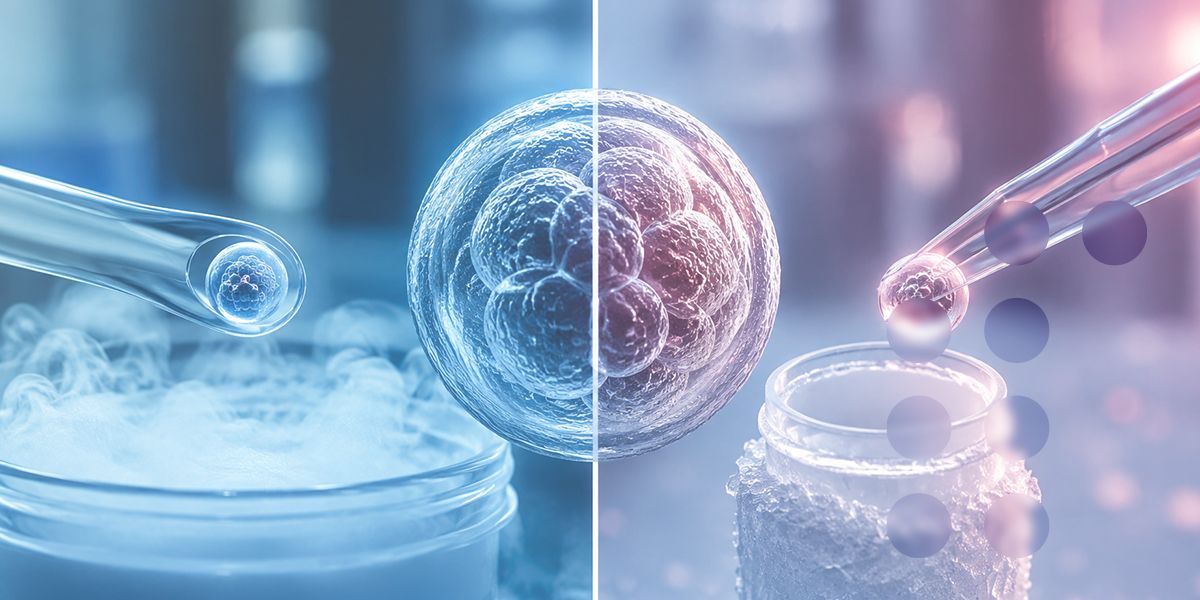

Vitrification vs. Slow Freezing: What Does the Scientific Evidence Show?
Cryopreservation has become an essential component of modern assisted reproductive technology (ART). Embryos and oocytes can be preserved for later use, allowing fertility treatment to be separated from the timing of ovarian stimulation, embryo creation, or other clinical circumstances.
For many years, conventional slow freezing was the established approach. Today, vitrification has become the dominant cryopreservation strategy for human oocytes and embryos in many IVF laboratories.
But is vitrification actually better than slow freezing? And, more importantly, does better survival in the laboratory translate into better clinical outcomes?
The scientific evidence largely supports vitrification, particularly for oocytes and embryos. However, the difference is not simply a matter of "freezing faster." Cryopreservation outcomes depend on cryoprotectant exposure, cooling and warming rates, sample volume, device design, embryologist competency, quality control, and the entire laboratory workflow.
This article reviews the underlying science and the evidence that matters in clinical practice.
What is the difference between vitrification and slow freezing?
Although both techniques are commonly described as "freezing," their physical mechanisms are different.
In conventional slow freezing, the sample is cooled gradually, typically at approximately −0.1 to −3°C per minute. The controlled reduction in temperature allows water to move out of the cell and promotes cellular dehydration while limiting intracellular ice formation. The sample is then stored in liquid nitrogen at approximately −196°C.
Vitrification takes a fundamentally different approach. Cells are exposed briefly to relatively high concentrations of cryoprotectants and then cooled extremely rapidly in a very small volume. Under appropriate conditions, the intracellular and extracellular solution transitions into a glass-like, amorphous state rather than forming damaging ice crystals. Modern rapid-cooling protocols can produce cooling rates of thousands of degrees Celsius per minute.
The key biological issue is therefore not simply temperature. It is ice.
Ice crystals can mechanically disrupt cellular structures and membranes. Vitrification is designed to minimize this risk by combining adequate dehydration, high cooling rates, very small sample volumes, and appropriate cryoprotectant concentrations.
There is also an important terminology point: ASRM notes that "slow freezing" is technically a misnomer because conventional protocols are more accurately described as slow cooling; ideally, intracellular ice should not form even during slow cooling.
Why does ice formation matter?
Oocytes and embryos contain substantial amounts of water. When water freezes, it can form ice crystals. The size, location, and dynamics of those crystals can influence whether cellular structures remain functional after warming.
For an oocyte, this is particularly important because the meiotic spindle and plasma membrane are sensitive structures. Damage during cryopreservation can affect survival and subsequent developmental competence.
For embryos, cryodamage can result in partial or complete cellular loss after warming. A blastocyst may survive, for example, but with a proportion of its cells damaged. The practical consequence is that an embryo may no longer be suitable for transfer or may have reduced developmental potential.
Vitrification seeks to avoid this crystallization process altogether.
However, vitrification introduces another challenge: the concentration of cryoprotectants is relatively high. Cryoprotectants can themselves be toxic if exposure is excessive or poorly controlled. Consequently, vitrification is highly dependent on accurate timing, temperature control, solution composition, sample volume, and operator technique.
What does the evidence show for embryos?
The evidence comparing vitrification with slow freezing is particularly strong for embryo cryopreservation.
An early systematic review and meta-analysis examined 8,824 cryopreserved human cleavage-stage embryos and blastocysts. Vitrification was associated with substantially higher post-warming survival than slow freezing. For cleavage-stage embryos, the reported odds ratio for survival was 15.57, while vitrified blastocysts also had significantly higher survival than slowly frozen blastocysts.
Later evidence has broadly supported this finding.
A systematic review and meta-analysis examining oocyte, embryo, and blastocyst cryopreservation found that pooled randomized controlled trial data from 3,615 embryos showed significantly better cryosurvival following vitrification compared with slow freezing, with a relative risk of 1.59 (95% CI 1.30–1.93). The authors concluded that available randomized evidence supported vitrification/warming over slow freezing/thawing, although they rated the quality of evidence for some clinical outcomes as low.
Randomized clinical studies have also demonstrated the practical difference.
In one randomized trial involving 1,798 cleavage-stage embryos, post-warming survival was 89.4% with one vitrification method and 87.6% with another, compared with 63.8% after slow freezing. The difference was statistically significant. Interestingly, implantation rates were not significantly different between the groups, demonstrating an important point: better laboratory survival does not automatically mean that every downstream clinical endpoint will improve.
Another randomized study of Day-3 embryos reported survival of 94.8% after vitrification compared with 88.7% following slow freezing, with higher subsequent blastocyst development in the vitrification group.
Most importantly, randomized clinical evidence has also reported an advantage in live birth. A randomized controlled trial published in Human Reproduction specifically evaluated live birth per embryo thawed/warmed after Day-3 embryo cryopreservation and concluded that vitrification produced a higher live-birth rate per warmed embryo than conventional slow freezing, attributable to better embryo survival, quality, and availability for transfer.
What about blastocysts?
Blastocyst cryopreservation is one of the areas in which vitrification has had a major practical impact.
Blastocysts contain a fluid-filled cavity and are therefore vulnerable to cryodamage and ice formation. Vitrification allows rapid transition to the glass-like state and, when appropriately performed, can produce very high survival after warming.
Population-based data have also been reassuring. A large cohort study comparing fresh, slow-frozen, and vitrified blastocyst transfers found that vitrified blastocyst transfer was associated with significantly higher clinical pregnancy and live-delivery rates than slow-frozen blastocyst transfer, while perinatal outcomes were comparable.
The evidence should nevertheless be interpreted correctly. Vitrification is not an independent guarantee of implantation or live birth. Embryo aneuploidy, embryo morphology, maternal age, endometrial preparation, uterine factors, sperm factors, embryo culture conditions, and many other variables influence the final clinical outcome.
Cryopreservation is one part of the ART chain.
What does the evidence show for oocytes?
The transition from slow freezing to vitrification has been particularly important for mature human oocytes.
Oocytes are more difficult to cryopreserve than embryos because of their large size, high water content, and sensitivity of the meiotic spindle.
Historically, slow cooling produced inconsistent oocyte survival. The development of rapid vitrification significantly improved outcomes.
A systematic review and meta-analysis of randomized controlled trials involving 4,282 vitrified oocytes, 3,524 fresh oocytes, and 361 slow-frozen oocytes found that oocyte survival was significantly higher after vitrification than slow freezing, with an odds ratio of 2.46. Fertilization was also higher after vitrification, with an odds ratio of 1.50.
A prospective randomized study similarly reported oocyte survival of 81% after vitrification/warming compared with 67% after slow freezing/thawing. Fertilization, cleavage, embryo morphology, and clinical pregnancy outcomes also favored vitrification in that study.
The American Society for Reproductive Medicine (ASRM) consequently identifies rapid-cooling vitrification as the standard approach for oocyte cryopreservation and notes that minimizing intracellular ice formation improves post-warming survival.
ESHRE likewise identifies oocyte vitrification as the method of choice for fertility preservation in relevant clinical settings.
There is, however, an important qualification: evidence is much less definitive for immature oocytes. A meta-analysis of germinal-vesicle-stage oocytes did not demonstrate a clear superiority of vitrification over slow freezing. Therefore, conclusions established for mature metaphase-II oocytes should not automatically be applied to every oocyte developmental stage.
Does higher survival mean higher pregnancy and live birth rates?
Not necessarily.
This is one of the most important points when interpreting cryopreservation studies.
Cryosurvival is a laboratory endpoint. Pregnancy and live birth are clinical endpoints.
A cryopreservation technique can produce a higher proportion of surviving embryos without producing an equally large improvement in implantation or live birth because many biological factors remain between warming and delivery.
Nevertheless, the overall direction of the evidence is favorable to vitrification.
The 2017 systematic review and meta-analysis concluded that vitrification/warming was superior to slow-freezing/thawing for cryosurvival of oocytes, cleavage-stage embryos, and blastocysts. It also found evidence suggesting improved clinical outcomes, although the authors emphasized that the quality of evidence for some clinical outcomes was lower than that for cryosurvival.
This distinction is important for laboratories and clinicians: the strongest and most consistent advantage of vitrification is its ability to preserve viable oocytes and embryos through the cryopreservation process.
Is vitrification safer for future pregnancies?
Current evidence does not indicate that properly performed vitrification creates a major additional risk to pregnancy or neonatal outcomes.
Studies evaluating vitrified embryos have generally reported reassuring pregnancy and neonatal outcomes. For example, research examining birth defects found no significant difference in the risk of birth defects between births following vitrified blastocyst transfers and vitrified cleavage-stage embryo transfers.
Longer storage also appears reassuring. A systematic review and meta-analysis involving more than 18,000 embryos found no significant differences in survival, miscarriage, live birth, or major malformation rates between embryos stored for more than 12 months and those stored for shorter periods after vitrification.
These findings support the clinical use of vitrification, but they should not be interpreted as proof that cryopreservation has zero risk. Long-term outcome research remains important, and clinical results depend on the entire ART process rather than cryopreservation alone.
What are the disadvantages of vitrification?
If vitrification is so effective, why does laboratory implementation require so much attention?
Because vitrification is technically demanding.
Slow freezing uses a programmed cooling curve and a relatively automated process. Vitrification depends more heavily on precise manual handling.
Critical variables include:
- Cryoprotectant exposure time
- Temperature of the solutions
- Equilibration and dehydration
- Sample volume
- Loading technique
- Cooling rate
- Warming rate
- Timing between steps
- Device characteristics
- Embryologist competency
- Liquid nitrogen storage conditions
- Quality-control procedures
ASRM emphasizes that vitrification outcomes are closely associated with operator skill and recommends structured quality control, competency assessment, monitoring of learning curves, and tracking of laboratory outcomes.
This is why two laboratories can use the same general vitrification principle and obtain different results.
The technology is only as reliable as the protocol and the laboratory implementing it.
Warming is as important as cooling
One of the most common misconceptions is that vitrification is primarily about ultra-fast cooling.
In reality, warming is equally critical and can be even more sensitive.
If a vitrified sample is warmed too slowly, ice formation or recrystallization can occur during the warming phase. This can undermine the very mechanism that vitrification was designed to achieve.
ASRM therefore emphasizes that the warming rate is at least as important as the cooling rate in successful vitrification.
For practical laboratory management, this means that a vitrification program should never be evaluated solely on its freezing step.
A complete assessment should include both:
Vitrification → storage → warming → post-warming assessment
The entire chain needs to be validated.
Open versus closed vitrification systems
Another practical consideration is the design of the cryopreservation device.
Vitrification systems may use open or closed configurations. Open systems can provide extremely rapid thermal transfer because the sample is directly exposed to liquid nitrogen. Closed systems provide an additional physical barrier between the specimen and liquid nitrogen.
The choice is therefore not simply a question of "which is faster?"
It involves balancing thermal performance, biological outcomes, contamination-control considerations, regulatory requirements, workflow, and laboratory validation.
A 2024 systematic review and network meta-analysis found no statistically significant difference in oocyte survival between open and closed vitrification systems in the analyzed evidence. However, the authors reported differences in some downstream developmental outcomes and suggested that closed systems may exert less detrimental effects on oocyte competence.
Earlier meta-analysis likewise found no significant difference in cryosurvival between open and closed systems in prospective oocyte studies.
Therefore, laboratories should select a validated system based on their specific clinical workflow, regulatory requirements, documented performance, and quality-management system rather than assuming that device configuration alone determines success.
What should an IVF laboratory measure?
A modern cryopreservation program should not be judged by a single percentage.
A useful quality dashboard should include, where applicable:
- Number of oocytes or embryos cryopreserved
- Cryopreservation rate
- Post-warming survival rate
- Complete versus partial survival
- Fertilization rate for warmed oocytes
- Cleavage and blastocyst development after warmed oocyte use
- Blastocyst re-expansion after warming
- Embryos suitable for transfer
- Implantation rate
- Clinical pregnancy rate
- Ongoing pregnancy rate
- Live birth rate
- Miscarriage rate
- Neonatal outcomes
- Device- or protocol-specific performance
- Operator-specific performance
- Deviations, incidents, and failed warming events
ASRM specifically recommends maintaining a database that tracks cryopreservation and warming outcomes through clinical and neonatal endpoints.
This approach transforms cryopreservation from a laboratory procedure into a measurable quality-management process.
What does this mean for embryologists?
For embryologists, the evidence supports several practical conclusions.
First, vitrification should be treated as a standardized laboratory procedure rather than an individual technique that can vary from operator to operator.
Second, competency must be demonstrated and monitored continuously. A new embryologist should not be considered fully competent merely because they have completed a training course. Their actual cryosurvival and clinical performance should be evaluated against validated laboratory benchmarks.
Third, timing is critical. Small deviations in exposure or handling can alter the osmotic and cryoprotective response of the cell.
Fourth, warming protocols deserve the same level of attention as vitrification protocols.
Fifth, every laboratory should validate its complete system—including solutions, devices, equipment, storage, warming protocol, documentation, and personnel—rather than evaluating individual components in isolation.
What does this mean for medical-device selection?
The shift toward vitrification has also increased the importance of cryopreservation consumables and devices.
A cryopreservation device should not be evaluated solely on its ability to hold an oocyte or embryo.
Relevant considerations include:
- Consistency of sample loading
- Minimal and reproducible sample volume
- Thermal performance
- Ease of handling
- Identification and traceability
- Compatibility with the laboratory workflow
- Reliability during warming
- Operator ergonomics
- Storage security
- Contamination-control strategy
- Regulatory status and documentation
- Lot-to-lot consistency
- Availability and quality of supporting protocols
A technically excellent cryopreservation medium cannot compensate for poor handling. Likewise, an excellent device cannot compensate for an inadequately validated protocol.
The best results come from an integrated system in which the consumable, protocol, equipment, storage process, and embryologist work together predictably.
Is slow freezing obsolete?
For human oocytes and embryos, slow freezing has largely been displaced by vitrification in contemporary ART because vitrification generally provides superior cryosurvival and, in several studies, improved clinical outcomes. ASRM recommends rapid-cooling vitrification as standard of care for cryopreservation of human oocytes and embryos.
The 2017 global-guidance-oriented systematic review reached a similar practical conclusion: laboratories that were still using slow freezing should consider transitioning to vitrification, particularly because of the improvement in cryosurvival.
However, "vitrification is better" should not become an excuse for oversimplification.
A validated slow-freezing program with strong outcomes may still be clinically functional, while a poorly implemented vitrification program can produce disappointing results.
The correct comparison is therefore not:
Slow freezing = old; vitrification = new.
It is:
Validated vitrification with competent operators and controlled warming generally provides better cryosurvival and is the preferred contemporary approach for human oocyte and embryo cryopreservation.
Vitrification vs. slow freezing: practical comparison
|
Factor |
Vitrification |
Conventional slow freezing |
|
Cooling |
Extremely rapid |
Controlled gradual cooling |
|
Physical state |
Glass-like, non-crystalline |
Controlled freezing with cellular dehydration |
|
Ice-crystal risk |
Minimized when properly performed |
Controlled but not eliminated |
|
Cryoprotectant concentration |
Relatively high |
Generally lower |
|
Sample volume |
Very small |
Generally larger |
|
Oocyte survival |
Generally higher |
Generally lower |
|
Embryo survival |
Generally higher |
Generally lower |
|
Operator dependence |
High |
Generally lower |
|
Warming sensitivity |
Extremely important |
Important |
|
Current role in oocyte cryopreservation |
Preferred/standard approach |
Largely replaced |
|
Current role in embryo cryopreservation |
Widely preferred |
Increasingly limited |
|
Quality control requirements |
High |
High |
The bottom line
The scientific evidence does not support treating vitrification as merely a newer version of slow freezing.
It represents a different cryobiological strategy.
For human oocytes and embryos, the evidence consistently shows that vitrification provides higher post-warming survival than conventional slow freezing. Randomized trials and systematic reviews support this advantage, and clinical studies indicate that the improved laboratory performance can translate into better pregnancy and, in some settings, live-birth outcomes.
For mature oocytes, vitrification has become the established approach because it substantially improves survival compared with conventional slow cooling.
For embryos and blastocysts, vitrification similarly provides higher cryosurvival and has become central to modern frozen embryo transfer programs.
But the final lesson is perhaps more important than the comparison itself:
Vitrification is not simply a cryopreservation method. It is a controlled laboratory system.
Its success depends on validated solutions, appropriate devices, precise timing, controlled temperatures, rapid and reproducible warming, competent embryologists, reliable cryostorage, traceability, and continuous quality monitoring.
For IVF laboratories seeking predictable outcomes, the objective should therefore not be to choose vitrification merely because it is the current standard. The objective should be to implement a complete, validated vitrification workflow that protects the biological material at every stage—from loading to storage to warming and eventual clinical use.
For medical-device manufacturers and suppliers supporting assisted reproduction, this creates an equally clear responsibility: cryopreservation products must be designed around reproducibility, usability, traceability, and the real-world requirements of embryology laboratories.
The scientific evidence has moved the field toward vitrification. The next challenge is ensuring that every laboratory can perform it consistently, safely, and to a high standard.
References
- American Society for Reproductive Medicine (ASRM). A Review of Best Practices of Rapid-Cooling Vitrification for Oocytes and Embryos: A Committee Opinion. Fertility and Sterility. 2021;115:305–310.
- Rienzi L, Gracia C, Maggiulli R, et al. Oocyte, embryo and blastocyst cryopreservation in ART: systematic review and meta-analysis comparing slow-freezing versus vitrification to produce evidence for the development of global guidance. Human Reproduction Update. 2017;23(2):139–155.
- Loutradi KE, Kolibianakis EM, Venetis CA, et al. Cryopreservation of human embryos by vitrification or slow freezing: a systematic review and meta-analysis. Fertility and Sterility. 2008;90(1):186–193.
- AbdelHafez F, Desai N, Abou-Setta AM, Falcone T, Goldfarb J. Slow freezing, vitrification and ultra-rapid freezing of human embryos: a systematic review and meta-analysis. Reproductive BioMedicine Online. 2010;20(2):209–222.
- Debrock S, Peeraer K, Fernandez Gallardo E, et al. Vitrification of cleavage stage day 3 embryos results in higher live birth rates than conventional slow freezing: a randomized controlled trial. Human Reproduction. 2015;30(8):1820–1830.
- Fasano G, Fontenelle N, Vannin AS, et al. A randomized controlled trial comparing two vitrification methods versus slow-freezing for cryopreservation of human cleavage stage embryos. Journal of Assisted Reproduction and Genetics. 2014;31:241–247.
- Balaban B, Urman B, Ata B, et al. A randomized controlled study of human Day 3 embryo cryopreservation by slow freezing or vitrification: vitrification is associated with higher survival, metabolism and blastocyst formation. Human Reproduction. 2008;23(9):1976–1982.
- Cobo A, Diaz C. Clinical application of oocyte vitrification: a systematic review and meta-analysis of randomized controlled trials. Fertility and Sterility. 2011;96(2):277–285.
- Smith GD, Serafini PC, Fioravanti J, et al. Prospective randomized comparison of human oocyte cryopreservation with slow-rate freezing or vitrification. Fertility and Sterility. 2010;94(6):2088–2095.
- Glujovsky D, Riestra B, Sueldo C, et al. Vitrification versus slow freezing for women undergoing oocyte cryopreservation. Cochrane Database of Systematic Reviews. 2014;CD010047.
- European Society of Human Reproduction and Embryology (ESHRE). ESHRE Guideline: Female Fertility Preservation. Human Reproduction Open. 2020;2020(4):hoaa052.
- Li Z, Wang YA, Ledger W, Edgar DH, Sullivan EA. Clinical outcomes following cryopreservation of blastocysts by vitrification or slow freezing: a population-based cohort study. Human Reproduction. 2014;29(12):2794–2801.
- Canosa S, Cimadomo D, Conforti A, et al. The effect of extended cryo-storage following vitrification on embryo competence: a systematic review and meta-analysis. Journal of Assisted Reproduction and Genetics. 2022;39:873–882.
- Pantos K, Maziotis E, Trypidi A, et al. The Effect of Open and Closed Oocyte Vitrification Systems on Embryo Development: A Systematic Review and Network Meta-Analysis. Journal of Clinical Medicine. 2024;13(9):2651.